What is VALVULAR DISEASE?
When blood is pumped through the heart, the flow of blood is in one direction only (blood flows into the heart and is pumped out of the heart).
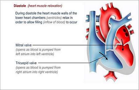
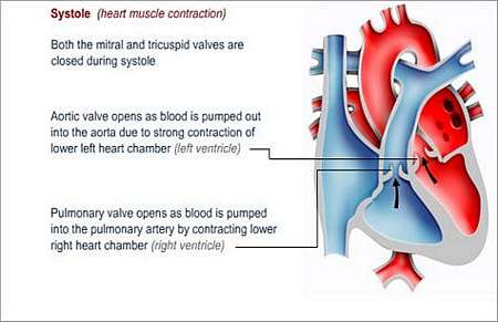
The heart valves play a critical role in the one-way movement of blood through the heart. With each heartbeat, there is contraction or dilatation of the heart muscle that results in pressure changes, which causes the heart valves to open and close like flaps.
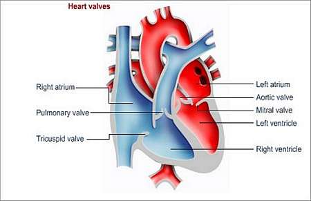
The heart has four valves
- Mitral valve
- Aortic valve
- Tricuspid valve, and
- Pulmonary valve
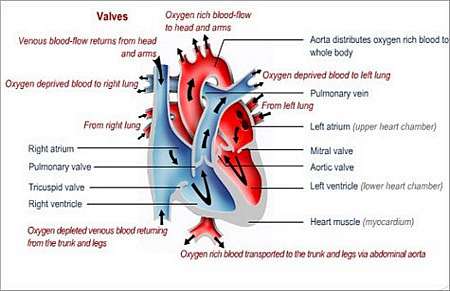
Deoxygenated blood returns from the body tissues and enters the right upper heart chamber (right atrium) from where it is pumped through the tricuspid valve into the right lower heart chamber (right ventricle).
Blood is then pumped (forced) through the pulmonary valve into the pulmonary arteries that transport the blood to the lungs where blood is oxygenated (mixed with oxygen).
From there it is transported to the left upper heart chamber (left atrium) from where it flows through the mitral valve into the left lower heart chamber (left ventricle).
The oxygen-rich blood is pumped through the aortic valve into the aorta that transports the blood via a progressively smaller branching network of arteries to all the tissues of the body.
There are two basic types of problems that can develop with valves. The valve can either become narrowed (stenotic) or it may not close properly, causing blood to regurgitate or seep backwards in relation to the normal flow (past the cusps - folds or flaps - of the incompetent valve).
Stenosis
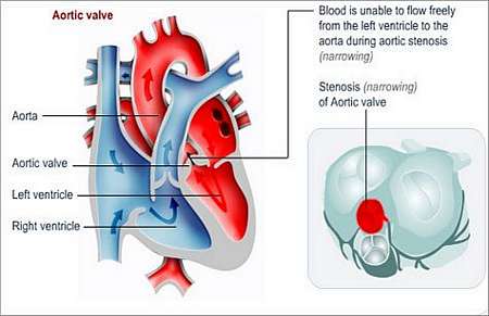
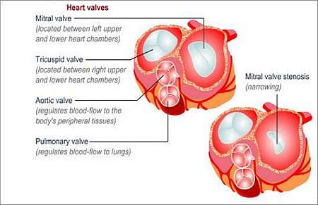
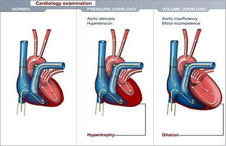
Stenosis occurs when the cusps (folds or flaps) of the valve are not able to open wide enough, causing the valve opening to remain small (narrow). This occurs when the valve cusps become stiff, when it thickens or when it fuses together. The heart muscle must work harder to force blood through the narrowed opening and this can cause heart failure.
Regurgitation
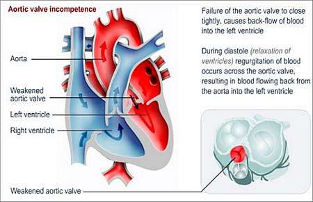
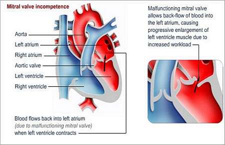
Regurgitation (incompetence or insufficiency) occurs when the valve does not close properly, allowing blood to leak backwards instead of moving in one direction only. If the condition worsens, the valve opening may become dilated and the heart muscle has to work progressively harder. Over time it causes the ventricle to enlarge and the muscle to undergo hypertrophy (get thicker) and less blood is pumped through the body.
Why does this happen and how may it affect the patients health?
- Calcific degeneration (mostly affects mitral and aortic valves; calcium build-up leads to thickened and ineffective valves)
- Abnormally shaped valves as a result of a birth defect (mostly affects aortic and mitral valves)
- Side effects of anti-obesity medicines, such as Redux and Fenphen
- Myxomatous degeneration (mostly affects mitral valve)
- Infective endocarditis (infection of the inner lining of the heart wall and valves)
- Coronary artery disease with secondary mitral regurgitation
- Rheumatic fever (before the days of antibiotics)
- Heart attack in combination with coronary artery disease
What symptoms may the patient experience?
Some patients do not experience symptoms at all. Symptoms depend on the severity of the valvular disease. Over time, patients with valvular disease will develop congestive heart failure. Other problems that may develop include arrhythmias (irregular heartbeat), blood clot formation and diseases of the heart muscle.
How is the diagnosis made and what special investigations are required?
- Clinical examination by a doctor
Valvular disease cause abnormal heart sounds, which can be heard on examination with a stethoscope.
- Echocardiogram
- Doppler ultrasound
Detects abnormal blood-flow through valves.
- Coronary angiogram
Assists to diagnose a narrowed valve or any backflow of blood.
- Chest X-ray
Can show if the heart is enlarged (might be due to an ineffective valve).
- Electrocardiogram (ECG)
By monitoring and charting the electrical impulse of the heart, abnormalities are detected and analysed.
- MRI (magnetic resonance imaging)
Allows three-dimensional visualization of the heart and itsinterventions valves.
What is the treatment?
- Observation and follow-up of asymptomatic patients.
- Lifestyle changes
Not much can be done to prevent valvular disease, but it is still important to live a heart-healthy lifestyle and control as many risk factors as possible. Rheumatic fever can be prevented by early diagnoses of throat infections. Any patient with a history of valvular disease must always take antibiotic medication before any dental or surgical procedure to reduce the risk of developing infective endocarditis.
- Medicines
Administered to ease the pain and other symptoms, to reduce the workload on the heart and to regulate the hearts rhythm.
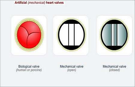
- Transcatheter interventions
Balloon valvuloplasty is a procedure that can be used to open narrowed valves. The procedure is performed in a cardiac catheterisation laboratory. A balloon-tipped catheter is inserted into the valve. When the balloon is inflated it enlarges the central area of the valves.
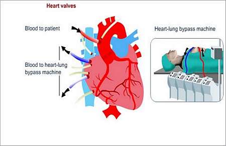
- Surgery
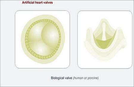
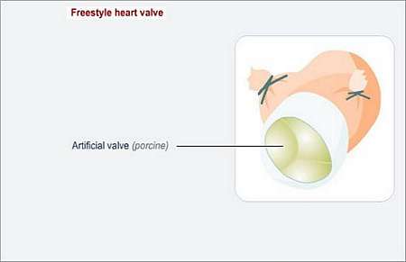
During valvular surgery, which is an open-heart procedure, valves may either be repaired or replaced. Surgeons use a heart-lung machine, because the heartbeat must be stopped for a short time during surgery.
<IMG src="http |