What is MAGNETIC RESONANCE IMAGING (MRI)?
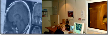
In this procedure, the patient is surrounded by a strong magnetic field. This enables the capturing of detailed pictures of the patients internal organs. Patients with pacemakers cannot undergo MRI scans, because it may interfere with the working mechanism of the pacemaker.
How is this done?
The patient lies supine in a short tunnel that holds a large magnet, while being exposed to short bursts of radio waves and magnetic fields. The patient has to lie absolutely still when the scan is performed. Images are created from the strong magnetic field and radio waves.

MRI scans can assist with making the following diagnoses
It can demonstrate abnormal heart function in diseases such as coronary heart disease (disease of the blood vessels that supply the heart muscle) and also cardiomyopathy (disease of the heart muscle itself).
It demonstrates and measures the flow of blood through major arteries.
It can also demonstrate structural defects of the heart.
© 2003 Prometheus™ Healthcare (Pty) Ltd
|