ECHOCARDIOGRAM (Cardiac ultrasound)
What is an Echocardiogram?
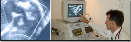
A pulse of high frequency sound is passed through the skin of the patients chest via a probe (recorder). In order for the probe to make good contact, lubricating jelly is rubbed on the skin of the chest. Echoes from various parts of the heart are reflected and picked up by the probe. These echoes are then displayed on an echocardiogram as a picture on a cathode ray screen.
As the probe is moved around on the chest wall, different parts of the heart can be viewed as images on the screen.
An echocardiogram provides information about the condition of the heart muscle and the valves of the heart. It is especially effective in children, because the procedure is not painful. It is often used for diagnosing birth defects of the valves or holes in the septum walls of the heart.
Other methods of performing echocardiography include
Doppler echocardiography
Measures the speed of the flow of blood in different parts of the heart; provides feedback on the status quo of heart valves.
Stress echocardiography
Carried out after the heart has been placed under stress by means of a drug or exercise.
Transoesophageal echocardiography
Allows detailed pictures of the heart to be taken via the oesophagus. The patient has to "swallow" a small probe, which is mounted at the end of a fine, flexible tube. Pictures are taken where after the tube and probe are gently removed.
© 2003 Prometheus™ Healthcare (Pty) Ltd
|